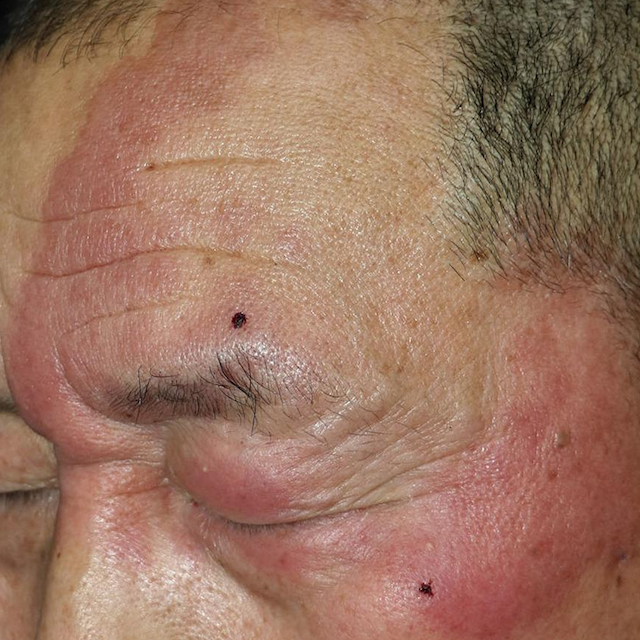
(BMJ)—A man in his 50s presented with a 6-year hx of an asymptomatic, well defined 12-cm diameter lesion that had been unresponsive to topical terbinafine. The patient had some loss of fine touch sensation and nociception, but peripheral nerves weren't palpable or tender. Acid fast bacilli in skin smears and results from nested PCR and DNA sequencing confirmed the dx. What is it?
mycosis fungoides
erythema annulare centrifugum
tinea faciei
borderline tuberculoid leprosy
You are correct. Borderline tuberculoid leprosy was diagnosed on the basis of clinical and lab findings. Recognizing that these clinical features may be caused by leprosy may enable earlier dx and tx before disability occurs. The erythema improved after a few weeks of multidrug tx for leprosy.

BMJ 2022;379:e071187
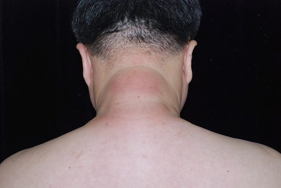
(BMJ)—This image shows a progressive, tight, erythematous, and hardened plaque that developed over 3 months on the neck of a man in his 40s. A skin biopsy sample showed thick collagen bundles with cleft-like spaces, without abnormalities in the epidermis. Colloidal iron staining showed mucin deposition among collagen bundles in the deep dermis. What’s the dx?
scleroedema diabeticorum
morphea
amyloidosis
cellulitis
You are correct. Scleroedema, a cutaneous mucinosis, can occur secondary to acute streptococcal throat infection, paraproteinaemia, or DM. The last of these, termed scleredema diabeticorum, is thought to be due to glycosylation of collagen in the dermis leading to an excessive accumulation of collagen and mucin. This patient, who had a raised hemoglobin A1c concentration of 8.9% and serum protein electrophoresis within reference limits, was treated with metformin for glycemic control. After 6 months both his hemoglobin A1c concentration and cutaneous lesions had improved.

BMJ 2024;384:e075940
By rray, 5 March, 2024
By rray, 22 February, 2024