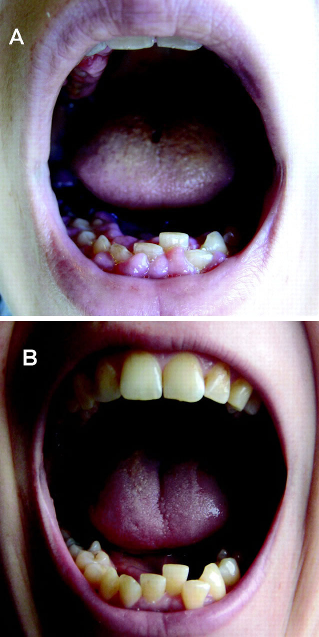
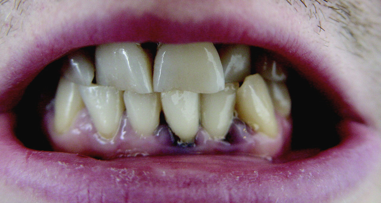
(BMJ) - A 35-yo pregnant woman developed gingival hyperplasia and lost 10 lbs over 5 wks w/ no other symptoms. Labs: leukocytosis, anemia, thrombocytopenia. Images show gums before and after resolution. What is the diagnosis?
Pregnancy-related gingival hypertrophy
HIV infection
Crohn disease
Infectious gingivitis
Acute myeloblastic leukemia
You are correct. This patient had acute myeloblastic leukemia confirmed by bone marrow aspiration. Pregnancy was terminated at around 22 weeks and chemotherapy promptly started. Her gingival hyperplasia improved within 5 days of starting chemotherapy (image A) and, by 51 days, had returned to normal without any odontological intervention (image B). Gingival hyperplasia is associated w/ acute myelomonocytic and monocytic leukemias and necessitates immediate investigation.
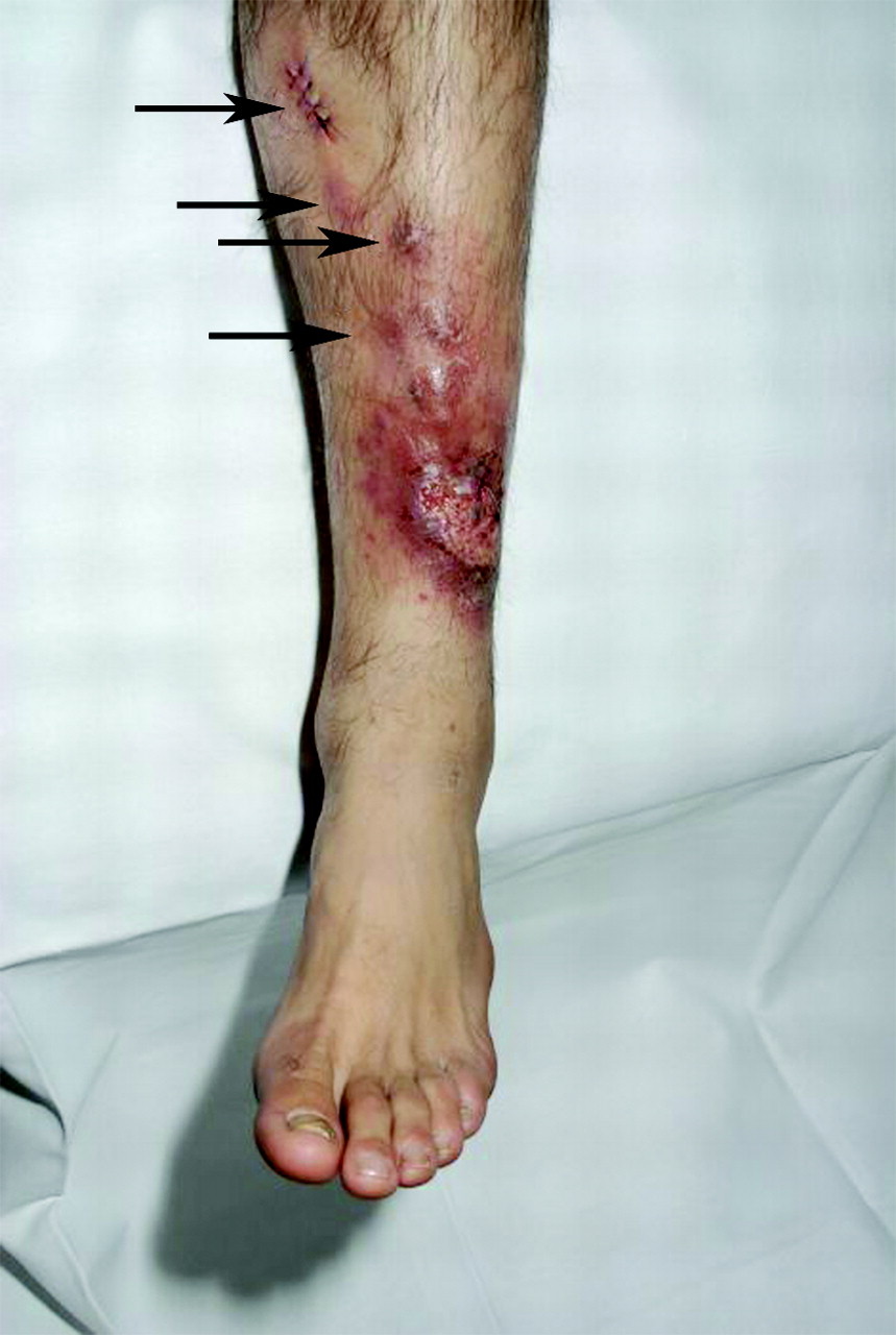
(BMJ) – A 40-yo man presented w/ a 6-wk hx of a mildly painful, enlarging ulcer on his lower leg after a trip to Afghanistan. Exam showed a large ulcer and proximal erythematous nodules. What is the diagnosis?
Sporotrichosis
Ulceroglandular tularemia
Cutaneous leishmaniasis
Leprosy
Pyogenic granuloma
You are correct. Cutaneous leishmaniasis due to Leishmania major (which is transmitted by sand fly bites) was confirmed by tissue polymerase chain reaction. This disease is seen frequently among returning military personnel. The patient was treated with intravenous sodium stibogluconate for 20 days.
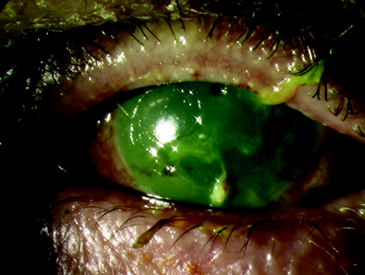
(BMJ) - A patient presented w/ eye complaints 36 hours after a car crash w/ airbag deployment. Fluorescein staining revealed diffuse uptake and corneal opacification. What is the diagnosis?
Corneal abrasion
Bacterial keratitis
Acute angle-closure glaucoma
Alkaline chemical burn
Ruptured globe
You are correct. This passenger sustained serious bilateral chemical injuries to the eyes due to airbag deployment. Airbags are inflated by nitrogen gas produced from sodium azide; deployment also produces sodium hydroxide, a highly alkaline and irritant chemical. This substance permeates through the airbag on deflation and can cause an alkaline burn to the eye. This pt sustained serious, potentially blinding corneal damage due to the late presentation. An eye exam should be considered in all pts exposed to airbag deployment.
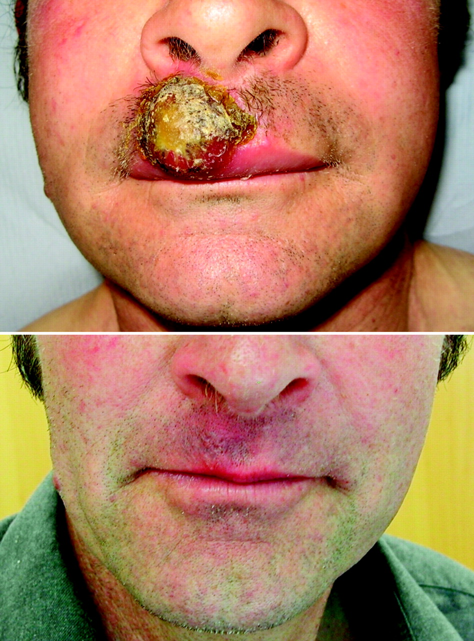
(BMJ) - A 56-yo sheep farmer presented w/ a lip lesion 3 wks after being butted on the mouth by a lamb. Biopsy showed no virus particles but confirmed the diagnosis. Images show lesion before and after tx w/ clobetasol propionate. What is it?
Pyogenic granuloma
Orf virus infection
Impetigo
Syphilis
Tick bite
You are correct. Pyogenic granuloma is a benign vascular entity often related to minor trauma. Lesions are usually painless but often bleed. Orf, a parapox virus infection associated w/ sheep, was excluded by lack of viral particles on bx. Treatment w/ topical super-potent corticosteroid ointment led to complete resolution over subsequent wks in this pt, obviating the need for surgery.
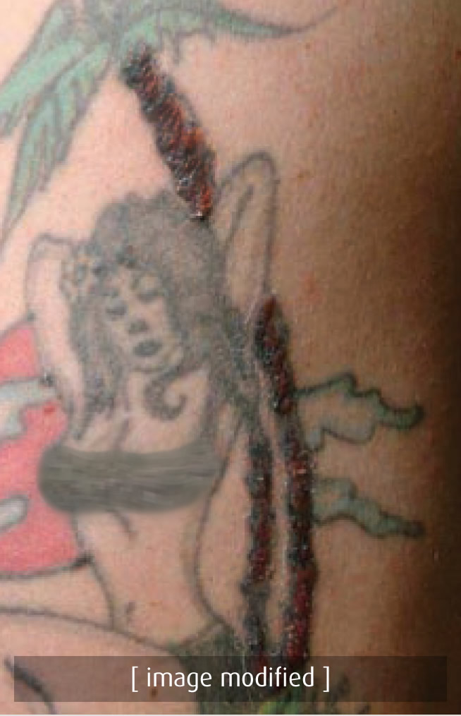
(BMJ) - A 40-yo man presented w/ polyarthralgia, chest pain, and malaise w/ tenderness and elevation of the “tree trunk” of his 20-yo tattoo. Chest CT showed hilar lymphadenopathy and interstitial changes. Skin bx confirmed the diagnosis. What is it?
Nontuberculous mycobacteria (NTM) infection
Tuberculosis
Histoplasmosis
Lymphoma
Sarcoidosis
You are correct. Tattoo reactions are a rare but recognized manifestation of sarcoidosis. In sarcoidosis, a state of granulomatous hypersensitivity exists, w/ reactions to different antigens in a range of organs. NTM infections due to contaminated tattoo inks have been reported, but present w/ localized skin, not pulmonary disease. The other choices are unlikely to be associated w/ tattoo reactions. Skin bx in this pt showed granulomatous infiltrate consistent w/ sarcoidosis. He responded well to a course of steroids.
(BMJ) - A 31-yo Romanian painter and decorator had a bluish line on his gums and iron-deficiency anemia w/ basophilic stippling on blood smear. A blood test confirmed the cause. What is the diagnosis?
Scurvy
Bismuth ingestion
Methamphetamine toxicity
Lead poisoning
Gingivitis
You are correct. A bluish line along the gingival margin (Burton line) is seen in chronic lead poisoning. An elevated serum lead level confirmed the diagnosis. Though lead paint is banned in many countries, safe paint removal practice remains unsatisfactory in many places, including Eastern Europe. Lead poisoning should thus be considered in migrant workers. Treatment is w/ chelation therapy.
(BMJ) - A 16-yo girl with history of atopic eczema developed multiple, painful, punched-out erosions over her forehead and eyelids after exposure to a person with cold sores. What is the diagnosis?
Coxsackie virus infection
Contact dermatitis
Bullous impetigo
Eczema herpeticum
Disseminated gonorrhea
You are correct. Eczema herpeticum typically presents with a "shotgun blast" appearance and is caused by herpes simplex virus. Patients with atopic eczema are susceptible to viral infections and should avoid contact with people who have active cold sores. Delay in treatment may cause scarring of skin and eyes and may be life-threatening in a child. This patient’s rash resolved rapidly with oral acyclovir.
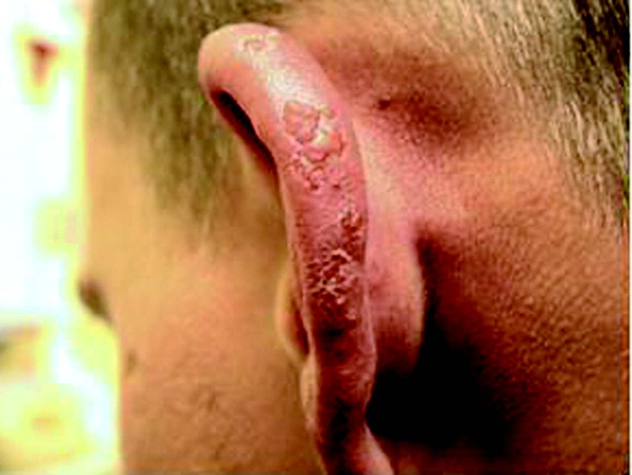
(BMJ) - A 38-yo sheep farmer had an itchy, painful, blistering eruption on his ear that occurred yearly at lambing time and resolved when lambing was over. Histology: heavy dermal perivascular lymphocytic infiltrate with normal overlying epidermis. What is the diagnosis?
Orf virus infection
Lupus erythematosus
Q fever
Lambing ear
Juvenile spring eruption
You are correct. Lambing ear is a recently identified ear rash that occurs in farmers during lambing season. This occupational disease occurs through close contact with products of conception. Juvenile spring eruption is a sun-induced skin condition affecting the light-exposed skin of the ears, most commonly in boys and young men in early spring. Orf virus is rarely transmitted to humans from sheep and causes papulopustular, viral-filled lesions. Q fever is a systemic illness that may also be transmitted by sheep. Cutaneous lupus erythematosus has distinct histology from lambing ear.
(BMJ) - A 37-yo man presented w/ swelling of the left ear for 10 days without hx of trauma. Antibiotics, aspiration, and I&D were ineffective and cultures of the aspirated fluid were negative. What is the diagnosis?
Relapsing polychondritis
Chondrodermatitis nodularis helicis
Auricular tuberculosis
Allergic dermatitis
Pseudocyst of the pinna
You are correct. Pseudocysts of the pinna do not respond to antibiotics, aspiration, or I&D; fluid simply reaccumulates. They are treated with a helical rim incision and removal of the anterior cyst wall. The posterior cartilage is curetted to remove any granulation tissue, and dental rolls are stitched to either side of the pinna to compress the area. Prompt treatment prevents deformity of the pinna.